


Motion Study to Improve PET Scan Quality
/As part of a project course, we, UBC Engineering Physics students Yas Oloumi, David Black, and Jeremy Wong, designed a breathing, humanoid phantom to study motion effects in Positron Emission Tomography (PET) in cooperation with Dr. Ivan Klyuzhin and Dr. Arman Rahmim at the Integrative Oncology Department of the BC Cancer Research Centre. With everything else figured out, questions remained about how to manufacture the lungs. We were lucky at this stage to come across Barber Prosthetics, and are very thankful for their help and guidance, which made this project a success.
Patient motion during PET scans can be a major detriment to image quality, leading to quantification errors and radiologists potentially missing malignant lesions. The primary source of movement in thoracic PET scans is respiratory, followed by cardiac motion. In order to study these problematic effects and potentially mitigate them through motion correction algorithms, anthropomorphic phantoms with accurate anatomy and realistic motion are needed. Anthropomorphic, or humanoid, phantoms are essentially model patients made of specific materials that simulate human tissue in their shape and their interactions with the gamma radiation of the PET scan. While anatomically accurate phantoms exist, none have been created with the realistic movement of a human, which makes them ineffective for motion studies. This is what we set out to do. The phantom can be scanned many times, unlike a real patient, and under precisely known and controlled conditions such as lung trajectory, breathing rate, and breathing tidal volume. This allows new scanning methods and algorithms to be developed and rigorously tested as was previously impossible, thus hopefully leading to better quantification and tumor detection in PET for the future.
Fig 1. Anatomically accurate phantom for PET imaging, with inserted lungs.
With the help of Malena, Daryl, and Dave at Barber Prosthetics, we were able to develop elastic lungs for such a phantom, which, together with a highly controllable, realistic breathing mechanism, are added to an existing, proven anatomically accurate phantom. Daryl, Dave, and Malena were instrumental to the lung manufacturing process, making multiple iterations of lung prototypes and helping us test to ensure an optimal design. The lung manufacturing process is outlined below:
A 3D lung model was extracted from a CT scan and simplified using mesh editing software.
From this model, a negative mold was 3D-printed in two halves out of polylactic acid (PLA). This is due to size constraints in 3D-printing.

Fig 2. Two-part, 3d-printed negative mold of the lungs used to create a dental plaster positive onto which the silicone elastomer is rolled.
3. Dental plaster was then poured into the negative mold using a hole at the base and allowed to set in the lung shape.

Fig 3. Dental plaster mold of the lungs.
4. Once the plaster dried, it was extracted from the mold and put in a polyvinyl alcohol (PVA) bag. This bag prevents the silicone catalyst from reacting with the plaster.
5. The silicone was then rolled onto the positive dental plaster mold of the lungs. In this critical step, it was important to ensure thin, very uniform wall thickness, which Daryl managed almost perfectly.
6. The silicone was reinforced and thickened at the base with an attachment point which could optionally be used to couple the lung to the respiratory mechanism to augment motion. It was also reinforced around the hole where the trachea were to connect.
7. The dental plaster with the rolled silicone was placed in an oven at 200°C for 12 hrs to cure the silicone.
8. The mold was removed from inside the silicone lungs through the breathing hole to which the trachea will be attached. This is possible because the material is highly flexible.
9. Through Barber Prosthetics’ excellent network, a machinist in the US fabricated custom fittings for us made entirely of plastic to connect the trachea securely to the lungs. The use of metal interferes with the creation of accurate PET images as the metal has a far higher extinction coefficient for 511KeV photons than anything in the body. Effectively, it blocks the emitted gamma radiation from reaching the sensors.

Fig 4. Silicone molded lungs (left) with custom fittings (right) for attaching trachea.
10. The fittings are screwed together with some silicone sealant, pinching the reinforced silicone around the breathing hole in the lung and creating an air-tight and robust seal.

Fig 5. Breathing tube connected to the lungs via the custom-made valve.

Fig 6. Lungs inside the rib cage and torso.
After some testing, we found that the actuated lung motion is quite realistic and consistent and will be very useful for its intended purpose. Some photos and videos which show the breathing can be seen here. As COVID-19 restrictions ease, we plan on verifying the phantom’s efficacy in PET and CT scans.
This was a fun, interesting, and impactful project which would have been impossible without the help of Barber Prosthetics. Their technical skill and creativity allowed Daryl, Dave, and Malena to bend their prosthetics expertise into creating realistic lungs with which innovative research on motion effects in PET can be carried out.

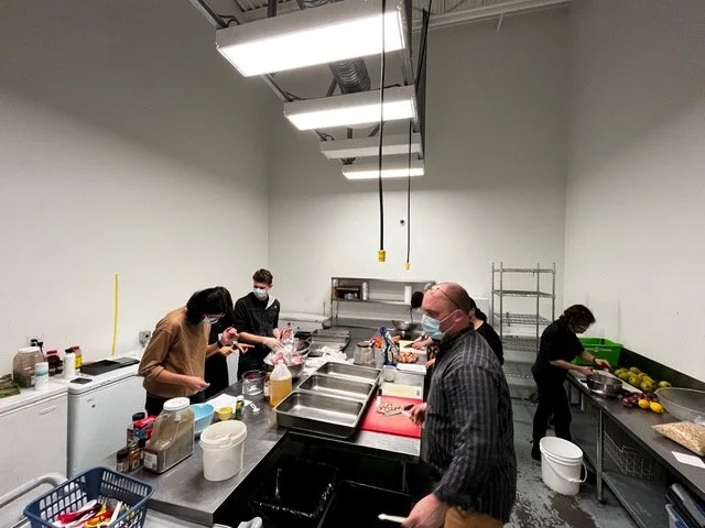
Fig 7. L – R: UBC Engineering Physics Students Yas Oloumi and David Black, with Barber Prosthetics staff Dave Moe and Daryl Murphy